Direct Connection of Myocarditis to Free-Floating S Spike Post mRNA Jabs - Study
A new study putting a finger on juvenile myocarditis causes post mRNA jabs.
Here’s a brief recap of the study “Circulating Spike Protein Detected in Post–COVID-19 mRNA Vaccine Myocarditis” (that was submitted on May 26, 2022, accepted on Nov. 23, 2022 and first published Jan. 3, 2023, so the jabbing may go on unhindered? ) by none other than Dr. Peter A. McCullough:
In particular, Dr. McCullough says:
The Spike protein they had evaded the apparently sufficient library of antibodies that were supposed to neutralize it. Thus, it is possible that some persons do not make specific neutralizing antibodies after injection, and thus, the Spike protein is able to circulate and damage the body, specifically the heart muscle.
Yet, I have to dissent. The study specifically tested for the presence of neutralizing antibodies and confirmed that, yes, the neutralizing antibodies are no different in those with myocarditis than in the healthy controls:
To assess whether this persistence of spike in patients with myocarditis was attributable to inadequate antibody neutralization, we carried out in vitro single-molecule array neutraliza-tion assays.20 We analyzed plasma samples from a subset of patients with myocarditis (n=9) and healthy vaccinated control subjects (n=14) for whom plasma samples were collected within 1 week after the sec-ond vaccine dose; however, no significant differences in antibody neutralization capacities were observed (Figure S6):
To return back to the study’s main finding:
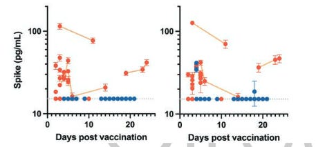
When analyzed according to time since vaccination, free S1, which was detected in only one patient with postvaccine myocarditis and one vaccinated control subject, was detected only within the first week. However, antibody-bound S1, which was detected in roughly one-third of both cohorts, could be detected up to 3 weeks after vaccination (Figure 4B). In contrast, both free and antibody-bound spike, which was detectable only in patients who developed vaccine-induced myocarditis, remained detectable up to 3 weeks after vaccination (Figure 4B). Longitudinal sampling displays a slow decline in both free and antibody-bound spike, suggesting that collecting blood at only a single time point was unlikely to miss circulating spike in healthy vaccinated control subjects.
Spend some time pondering Figure 4B, and 4A, for good measure, to understand what is going on.
The article differentiates between the full S spike and the S1 subdomain of the S spike. Here’s what that means:
Structure of the S protein
With a size of 180–200 kDa, the S protein consists of an extracellular N-terminus, a transmembrane (TM) domain anchored in the viral membrane, and a short intracellular C-terminal segment [11]. S normally exists in a metastable, prefusion conformation; once the virus interacts with the host cell, extensive structural rearrangement of the S protein occurs, allowing the virus to fuse with the host cell membrane. The spikes are coated with polysaccharide molecules to camouflage them, evading surveillance of the host immune system during entry [12].
The total length of SARS-CoV-2 S is 1273 aa and consists of a signal peptide (amino acids 1–13) located at the N-terminus, the S1 subunit (14–685 residues), and the S2 subunit (686–1273 residues); the last two regions are responsible for receptor binding and membrane fusion, respectively. In the S1 subunit, there is an N-terminal domain (14–305 residues) and a receptor-binding domain (RBD, 319–541 residues); the fusion peptide (FP) (788–806 residues), heptapeptide repeat sequence 1 (HR1) (912–984 residues), HR2 (1163–1213 residues), TM domain (1213–1237 residues), and cytoplasm domain (1237–1273 residues) comprise the S2 subunit (Fig. 2a) [13]. S protein trimers visually form a characteristic bulbous, crown-like halo surrounding the viral particle (Fig. 1a).
In the native state, the CoV S protein exists as an inactive precursor. During viral infection, target cell proteases activate the S protein by cleaving it into S1 and S2 subunits [17], which is necessary for activating the membrane fusion domain after viral entry into target cells [18]. Similar to other coronaviruses, the S protein of SARS-CoV-2 is cleaved into S1 and S2 subunits by cellular proteases
That is, the S1 subunit is cleaved (split) from the rest of the S spike in the process of the virus infecting a human cell. But as there is no virus post-jab, the S spike alone is circulating in the jab recipient’s bloodstream. So what the study is telling us, is that there is either a physiological difference between the myocarditis sufferers and the controls, or that the S spikes are somehow different. The latter is likely, as we know that the synthetic S spike is being produces in human cells, post jab, with many translational errors due to the mRNA “optimizations” undertaken by jab purveyors to make said mRNA code both more stable and faster to translate. Another possibility is that the mRNA in the jab itself is “messed up”, as EMA noticed in Dec. 2020 that up to half of the mRNA in Pfizer jabs was not that of the promised S spike mRNA (in my post “Zeroing in on Gifts from “Science” to Humanity”, Nov. 6, 2021).
I personally suspect that it’s the slight variance in the mRNA code that produces more of the uncleaveable S spike in the myocarditis victims. Or, as the reader IPA points out: “Another issue might be how much of the poison was injected directly into the blood stream.”
Also, you can see in Figure 4B that the amount of total antigen (S Spike) and free antigen is actually increasing in one myocarditis subject well after 20 days post jab.
The study makes an effort to dissuade us from thinking that S spike is the actual cause of myocarditis (bad for business). I leave you with this presentation by Dr. Sukrit Bhakdi:
Here are some additional points from Jikkyleaks’ tweets:


It could be just a question of time. Another issue might be how much of the poison was injected directly into the blood stream.
This presentation by Dr. Bhakdi is excellent immunology 101. My take is that there victims got a jab which did as designed and the healthy controls were the lucky ones. That is Sasha Latypova's take with these DOD prototypes. https://open.substack.com/pub/sashalatypova/p/nobody-knows-what-is-in-the-vials?utm_source=direct&r=pbkzb&utm_campaign=post&utm_medium=web